Molecular Fluorescence: A New Signature for Biological Imaging

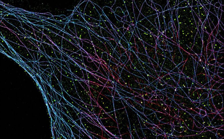
Observing multiple types of proteins simultaneously, within the thickness of a cell and on the scale of a few nanometers, remains one of the major challenges in bioimaging. Super-resolution fluorescence microscopy has already made it possible to overcome the limit imposed by light diffraction. But when it comes to tracking multiple biological targets simultaneously, current methods rely primarily on the color of fluorophores. However, their spectra overlap—much like shades that are too close together on a palette—which limits the number of distinguishable proteins.
A new approach, called Brightness Demixing, proposes a different way of looking at these fluorescent molecules. Rather than identifying them by their color, it exploits their brightness: the amount of light emitted by each individual molecule. This brightness depends on the fluorophore’s ability to absorb light and then re-emit it. It thus becomes a measurable signature, comparable to the intrinsic brightness of a star in a dark sky.
To obtain this information, researchers conduct a detailed analysis of the blinking events of fluorescent molecules. By taking multiple measurements, they quantify the flux of emitted photons and can classify fluorophores even when they are observed simultaneously in the same detection channel. The method therefore requires neither an additional camera nor complex spectral separation, and remains compatible with the localization microscopes already in use.
The team demonstrated this strategy for imaging two and then three biological targets, both in two dimensions and in three dimensions. Complex cellular structures, including nuclear pores, tubulin, and clathrin in COS-7 cells, served as test cases. By using a single excitation wavelength, Brightness Demixing also minimizes chromatic aberrations that can blur the superimposition of images obtained using multiple colors.
This method does not eliminate all the limitations of super-resolution imaging: it requires fluorophores with sufficiently distinct intensities and rigorous signal analysis. However, it adds a simple yet powerful dimension of information to single-molecule microscopy. Ultimately, it could facilitate the study of the nanoscale architecture of cells and help us better understand the organization of proteins involved in biological and pathological mechanisms.
Notes
This research was conducted by researchers from Institut Langevin (ESPCI Paris – PSL, CNRS) and the Institut des Sciences Moléculaires d’Orsay (CNRS, Université Paris-Saclay). It received support from the AXA Research Fund, the Labex WIFI programme, the ANR TimeLoc project and the ERC TimeNanolive project.
References
Le, L., Sreenivas, S. K., Fort, E. et al. “Brightness demixing for simultaneous multi-target imaging in 3D single-molecule localization microscopy.” Nature Methods (2026).
https://doi.org/10.1038/s41592-026-03118-6
 Published on 07/06/2026
 Research
 The Langevin Institute
 Emmanuel Fort
 Paul Turpault, paul.turpault@espci.fr