Pink October & Breast Cancer: What New Images May Reveal
Better characterizing a lesion, examining the structure of a tumor, and identifying its boundaries during surgery: imaging plays a role in several stages of breast cancer diagnosis and treatment. To mark Pink October, we shine a spotlight on three approaches being studied in the laboratories at ESPCI Paris–PSL, in collaboration with medical teams.

A medical image provides information about the shape and location of a lesion. But it can also reveal less visible characteristics: tissue stiffness, tissue structure, or the blood vessels running through the tissue. Can this information help distinguish a benign lesion from a malignant tumor? Can it help guide a biopsy or surgical procedure more effectively?
These issues bring together physicists, image processing specialists, and physicians. Their work aims to enhance diagnostic tools, with a common goal: to determine what information is truly useful for medical decision-making.
Cross-referencing data to better characterize tumors
At the Paris Institute of Physics for Medicine (Inserm, CNRS, ESPCI Paris – PSL), Béatrice Berthon is leading the MUSCAD project, which is supported by an ERC Starting Grant and will begin in 2025 for a duration of five years.
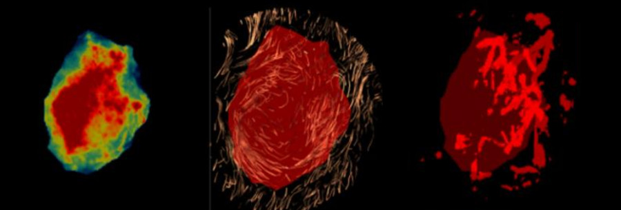
The goal is to gather, in a single three-dimensional ultrasound scan, various types of information about a tumor: its mechanical properties, its vascularization, and the organization of its tissues. Machine learning methods will be used to analyze this data and evaluate its ability to characterize breast tumors.
This approach builds on research into shear-wave elastography, to which Mathias Fink, Mickaël Tanter, and their teams at ESPCI have contributed. Already in clinical use, this technique measures tissue stiffness and provides information that complements ultrasound imaging.
With MUSCAD, the goal is to go a step further by cross-referencing multiple indicators. The clinical studies planned with the Institut Curie will assess the value of this approach, with the aim of eventually reducing the number of unnecessary biopsies.
Exploring Breast Tissue Using the Speed of Sound
At the Langevin Institute (CNRS, ESPCI Paris – PSL), Alexandre Aubry and Baptiste Hériard-Dureuil are exploring another property: the speed at which sound travels through breast tissue.
Their DIONISOS project, supported by a 2025 ERC Proof of Concept grant, aims to develop a method for imaging breast tumors based on this measurement. The goal is to investigate whether variations in sound speed can provide additional information for characterizing the malignancy of lesions.
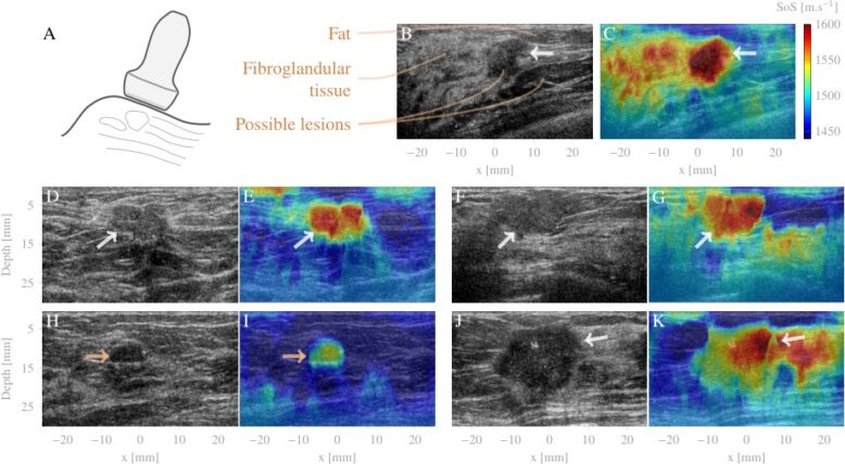
This study highlights a central question in imaging research: beyond the visual quality of an image, what physical quantities should be measured to better understand the tissue being observed?
Analyzing tissue during surgery
When a tumor is removed, it is essential to accurately identify its boundaries in order to preserve healthy tissue while removing all cancerous tissue.
In 2023, a preclinical study involving the Langevin Institute, Gustave Roussy, and the company then known as Aquyre Biosciences explored a method for the rapid analysis of surgical breast specimens. This research, to which Olivier Thouvenin contributed, combined optical microscopy without prior staining and machine learning to identify cancerous tissue.
Based on 51 samples, this study demonstrated the potential of this approach to better identify tumor margins during surgery and, ultimately, reduce the need for repeat surgeries. However, these results called for validation using a larger and more diverse set of samples.
 Published on 26/10/2026
 Research